2D/3D GALLERY
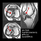
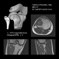
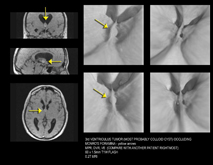
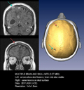
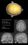
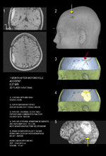
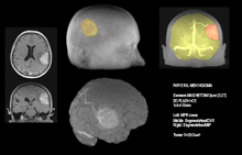
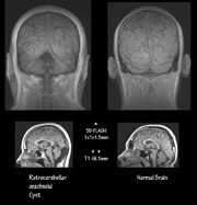
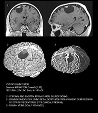
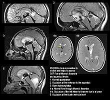
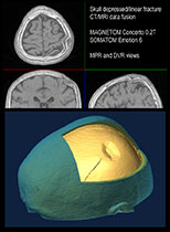
Once an object being tomographed is cut and examined slice by slice we are now ready to make a second logical step and reassemble our object in order to evaluate it as a whole with pathology accentuated and easy for visual perception. That's what a computer can do by means of a technique called "three-dimensional reconstruction". Until recently the domain of volumetric imaging remained a property of dedicated and quite expensive UNIX-based platforms featuring RISC-type processors and proprietary software. Now a modern high-performance personal computer is successfully used for the same task at a fraction of cost of its predecessors! A variety of Windows software suitable for medical 3D visualization and postprocessing are also available, free or commercial. Here are some examples done on a PC/Windows "workstation":

