ANATOMY AND PHYSIOLOGY
In order to appreciate our fertility we must understand the body's reproductive system, which enables men and women to become parents. Our discussion of its Anatomy and Physiology will therefore include:
a description of the male and female organs of reproduction (Anatomy)
an explanation of the function of the male and female organs of reproduction (Physiology)
an introduction of the concept of "combined fertility", the way by which the reproductive systems of men and women complement each other and work together to bring new persons into existence.
|
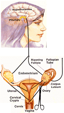
FEMALE REPRODUCTIVE ANATOMY |
The uterus is the pear-shaped and - sized muscular organ, designed to nurture the fertilized egg cell. It can expand to many times its size.
The endometrium is the lining of the uterus that, under the influence of the reproductive hormones, is prepared each cycle for the implantation of the fertilized egg cell.
ANATOMY AND PHYSIOLOGY
In order to appreciate our fertility we must understand the body's reproductive system, which enables men and women to become parents. Our discussion of its Anatomy and Physiology will therefore include:
a description of the male and female organs of reproduction (Anatomy)
an explanation of the function of the male and female organs of reproduction (Physiology)
an introduction of the concept of "combined fertility", the way by which the reproductive systems of men and women complement each other and work together to bring new persons into existence.
|
FEMALE REPRODUCTIVE ANATOMY |
The uterus is the pear-shaped and - sized muscular organ, designed to nurture the fertilized egg cell. It can expand to many times its size.
The endometrium is the lining of the uterus that, under the influence of the
reproductive hormones, is prepared each cycle for the implantation of the
fertilized egg cell.
The Fallopian tubes are the tubes which extend from each side of the uterus, and
provide the passageway for the egg cell as it migrates from the ovary to the
uterus. The trumpet-shaped ends of the Fallopian tubes are lined with millions
of tiny
feathery fingers that are constantly in motion to draw the egg cell into the
tube once it leaves the ovary. Fertilization occurs in the outer third of the
Fallopian tube. The fertilized egg cell will then reach the uterus in about six
days when it implants in the endometrium.
The ovaries are the almond-shaped female sex glands located on each side of the uterus, near the open end of each Fallopian tube. The pecan-sized ovaries produce egg cells or ova, and the hormones estrogen and progesterone. An infant girl's ovaries contain roughly 400,000 immature follicles, the structures within the ovary which consist of a layer of cells surrounding a fluid-filled cavity in which the immature egg cell is located.
Only about four hundred egg cells will actually be released throughout a woman's reproductive years, usually one per cycle for approximately thirty years.
The cervix is the narrow neck of the lower end of the uterus that connects the main body of the uterus with the vagina, and through which sperm enter the uterus and babies are born. It is like a valve that opens and closes. The cervix contains glands that produce mucus at various times during the fertility cycle.
The vagina is the muscular tube that connects the uterus with the outside of the body. It is the female organ of sexual intercourse - it receives the seminal fluid containing the sperm. The vagina serves as the birth canal - a passageway for the baby during birth.
The egg, or ovum is the female reproductive cell - the "seed of new life." It is the largest cell in the female body. The egg contains twenty-three chromosomes and is contained in follicles inside the ovary. Usually one egg matures per cycle and is released in the process of ovulation. The egg lives about twelve to twenty-four hours after leaving the ovary.
|
FEMALE REPRODUCTIVE PHYSIOLOGY |
External
The vulva is comprised of the external sex organs - the labia majora, labia minora and clitoris.
The reproductive process is controlled by a special part of the brain called the Hypothalamus. It does this by controlling the production of reproductive hormones secreted by the pituitary gland.
The Pituitary is a pea-sized gland located just below the hypothalamus. It secretes a number of hormones, but two in particular act directly on the ovaries. These two hormones are called follicle stimulating hormone (FSH) and luteinizing hormone (LH). Once the hypothalamus has initiated a woman's cycling mechanism, she will always be in a cycle unless she is pregnant or postmenopausal.
Each new cycle begins about two or three days ahead of menstruation, when the hypothalamus sends a releasing hormone to the pituitary to begin producing follicle stimulating hormone (FSH).
FSH is carried in the bloodstream and stimulates a group of follicles in the ovary to begin maturing.
As the follicles mature, they produce estrogen.
|
FEMALE ANATOMY AND PHYSIOLOGY |
Estrogen stimulates the cervix to begin to produce fertile-type mucus and the
lining of the uterus to thicken in preparation for the fertilized egg cell.
During puberty, an increase in estrogen causes secondary characteristics to
appear, such as the growth of body hair, breast enlargement, contouring of the
hips, and so on.
The intricate feedback control of hormones between the hypothalamus, pituitary
and ovaries, soon causes one follicle to become dominant and the others to
subside and shrink away; it also causes the release of a surge of the second
pituitary hormone, LH.
The surge of LH is the essential trigger for ovulation, causing final growth and
maturation of the egg cell, ovulation, and the formation of the corpus luteum
from the now empty follicle.
The corpus luteum, stimulated by LH, begins to secrete progesterone and
estrogen. The rising level of progesterone causes an abrupt change in the
cervical mucus, from wet and slippery to sticky, or perhaps, causing it to
disappear altogether. Progesterone also causes the woman's resting body
temperature to rise slightly.
The uterus, also stimulated by the estrogen and progesterone secreted by the
corpus luteum, continues to prepare for the fertilized egg cell.
The secretory capacity of the corpus luteum is maintained by LH. If
fertilization does not occur, the progesterone secreted by the corpus luteum
inhibits the production of FSH. This results in a fall in LH secretion by the
pituitary and consequently, a cessation of secretory activity by the corpus
luteum, usually 9-11 days after ovulation.
When the corpus luteum regresses, estrogen and progesterone levels fall rapidly.
The thickened lining of the uterus is no longer maintained and is sloughed off
as menstruation, ending the cycle.
During breast-feeding, the brain temporarily stops the activity of the ovaries
in order to protect the body's ability to provide nourishment to the baby. This
helps the baby to develop better both physically and emotionally. The mother's
defenses against infection are passed on to the baby through her milk. As
pediatrician and author, Dr. Robert L. Jackson writes:
| [E]cological breast-feeding is the natural and safe, but not well-appreciated mode of child spacing. We have gradually learned to document the hormonal changes that automatically result during breast-feeding in a period or space of about two years between births. The ovulation suppressant effect of ecological breast-feeding with consequent lactational amenorrhea...is now a well-recognized and proven fact. |
|
MALE REPRODUCTIVE ANATOMY |
The reproductive process in the male is also controlled by the hypothalmus. It does this by controlling the production of reproductive hormones secreted by the pituitary gland.
The pituitary gland is the pea-sized gland located just below the hypothalmus. It secretes a number of hormones, two of which, FSH and LH, control the male reproductive functions.
|
MALE REPRODUCTIVE ANATOMY |
The reproductive process in the male is also controlled by the hypothalmus. It does this by controlling the production of reproductive hormones secreted by the pituitary gland.
The pituitary gland is the pea-sized gland located just below the hypothalmus.
It secretes a number of hormones, two of which, FSH and LH, control the male
reproductive functions.
The testes (testicles) are the two male sex glands in which sperm and testosterone, the primary male hormone, are produced. The testes are located outside the abdomina-pelvic cavity in a pouch called the scrotum. They are located externally instead of internally because of the lower-than-normal body temperature required to produce sperm. Under the stimulus of FSH, sperm are produced, and under the stimulus of LH, the Leydig cells secrete androgens (male sex hormones).
The scrotum is the external pouch suspended behind the base of the penis, containing the testes.
The epididymis is the long coiled tube inside the testicles that carries sperm from the testes to the vas deferens. Sperm are collected and stored here as they leave the testicles. They also continue to mature here.
The vas deferens, commonly called the sperm duct, is a long fibromuscular tube that transports sperm from the epididymis to the urethra.
The prostate gland is one of the male accessory sex glands which surrounds the lower part of the bladder and the upper urethra. Its secretion is part of the seminal fluid.
The seminal vesicles, a pair of sac-like structures located behind the prostate, are associated with the vas deferens and secrete a thick, yellowish, alkaline fluid which forms the bulk of the semen.
The urethra is a narrow canal which, in women, extends from the bladder to the vulva; in men, the urethra passes through the penis and opens at its extremity, the glands of the penis. In men and women the urethra conveys urine from the bladder to the exterior of the body; in men, it also conveys the semen (but not at the same time).
The penis is the male genital organ, the organ used during sexual intercourse. It ejaculates the seminal fluid containing the sperm. It is located in front of the scrotum.
Ejaculation is the discharge of semen from the penis. Impulses from the spinal cord centers cause contractions of the epididymis and vas deferens, which expel sperm into the urethra. Two to four cubic centimeters of semen, containing approximately 300 million sperm, are expelled at each ejaculation.
The sperm is the male reproductive cell - the "seed of new life." It is the smallest cell in the male body and is continuously produced by the testicles from puberty onward. A sperm cell contains twenty-three chromosomes, including the X or Y chromosome that determines gender.
|
Combined Fertility and Conception |
Listing the similarities and differences between male and female Anatomy and Physiology helps us to recall what we have learned and, at the same time, helps us appreciate the gift of combined fertility.
Similarites
fertility begins to mature at puberty
increase in hormones triggers puberty
pituitary gland controls the reproductive system through hormones
sex cells have twenty-three chromosomes
| Male | Female |
| • Sperm - reproductive cell • Sperm - smallest cell in body • Testicle - sex gland • Penis - organ of sexual intercourse • Sperm production begins at puberty • Testosterone - primary hormone • Continuously fertile after onset of puberty |
• Ovum (egg) - reproductive
cell • Ovum - largest cell in body • Ovary - sex gland • Vagina - organ of sexual intercourse • Eggs present in ovaries at birth • Estrogen and progesterone - primary hormones • Fertile only for a few days each cycle |
The similarities of male and female Anatomy and
Physiology make the two systems compatible; differences between the two make
them complementary and allow them to combine with each other in the miracle of
bringing into existence a new human being. Combined fertility is the power of a
man and woman to participate in the creation of a human life. Three things are
needed in order for conception to take place, for a sperm to penetrate the egg
at fertilization: sperm, ovum and cervical mucus.
After ovulation and for most of the cycle, the cervix is completely closed by a
thick, sticky mucus which helps protect a woman's system from infection by both
its physical and antibiotic properties and prevents sperm from entering the
uterus. When sperm are kept out of the uterus, they have a very short life span
in the hostile environment of the vagina.
Close to ovulation, however, this mucus plug is released and the cervix begins
to produce a different kind of mucus which provides the ideal environment for
sperm. This mucus protects and nourishes the sperm, filters the seminal fluid so
that the abnormal sperm are eliminated, and provides channels through which the
remaining sperm are able to enter the uterus.
About three hundred million sperm are ejaculated and deposited in the vagina
during an act of intercourse. The fertile cervical mucus helps the sperm survive
the journey to meet the egg in the Fallopian tube. It filters out abnormal sperm
and enables the healthy ones to enter the cervix through channels or
passageways, as if swimming up a river. The mucus also nourishes the sperm so
they will be full of energy and vitality when they reach the egg in the tube. In
addition, the mucus keeps some sperm alive and healthy for three to five days in
the cervical crypts (tiny "niches" in the cervical wall).
The surviving sperm must travel the length of the uterus as they make their way
toward the tubes. The sperm cannot know whether the egg is to the right or the
left, so they enter both tubes.
Of the millions of sperm to enter the vagina, only a few hundred will actually
reach the Fallopian tubes and only one will penetrate and fertilize the egg.
Conception occurs as the sperm and egg unite and new human life begins. The
hereditary characteristics of the parents that are contained in the sperm and
egg mingle and are passed on to their child. At this moment, the new person's
genetic makeup--the color of his eyes and hair, and so forth is determined. He
is unique and unrepeatable. Dr. Jerome Lejeune explains:
| [L] ife has a very long history, but each of us has a unique beginning, the moment of conception. We know and all the genetics and all the zoology are there to tell us that there is a link between the parents and the children...As soon as the twenty-three chromosomes carried by the sperm encounter the twenty-three chromosomes carried by the ovum, the whole information necessary and sufficient to spell out all the characteristics of the new being is gathered... In the same way, when the information carried by the sperm and by the ovum have encountered each other, then a new human being is defined because its own personal and human constitution is entirely spelled out. There exist alot of minute differences in the message given by the father and the one given by the mother, even by the same person; we do not give exactly the same minute information in each sperm or in each egg. It follows that the voting process of the fertil- ization produces a personal constitution, entirely typical of this very one human being, which has never occurred before and will never occur again... Now, chromosomes are a long thread of DNA in which information is written. They are coiled very tightly on the chromosomes, and, in fact, a chromosome is very comparable to a mini-cassette, in which a symphony is written, the symphony of life. Immediately after the moment of conception, when the egg and sperm unite, the cells of the new human being begin to divide and multiply at an incredibly fast rate. The new, unique life now moves down the Fallopian tube to implant in the lining of the uterus, where it is surrounded by the cells of the endometrium (which provides protection and nourishment). Meanwhile, at the cervix, the mucus plug is being formed that will help protect the developing baby until it is time for birth. The baby will grow in the uterus for the next nine months until, at the start of labor, the mucus plug is expelled and he is born. |