
The Mechanical Assessment of Low Back Pain Patients with Painful
Lumbar Facet Joints
Sharon Young, PT, Cert MDT
The use of fluoroscopically-guided diagnostic injections is currently regarded as the most reliable means of identifying symptomatic lumbar discs, lumbar facet joints and sacroiliac joints. Each of these structures can be hydraulically distended to provoke a concordant (familiar) pain response when symptomatic. Subsequent introduction of an anesthetic with substantial pain relief fulfills the criteria for identifying a symptomatic structure by diagnostic injection.
Identifying symptomatic structures by physical examination is not so clear cut. The centralization phenomenon has been shown to be predictive of discogenic pain1. The source of pain which only peripheralizes during dynamic mechanical testing can be discogenic1, but cannot always be readily determined. Some examples of pathologies present in patients whose pain only peripheralized during testing includes a gas filled disc, a swollen dorsal root ganglion and an avulsion ridge of the vertebral end plate11.
It has been found that use of the McKenzie lumbar examination augmented by specific sacroiliac joint (SIJ) pain provocation tests4 can reliably predict painful sacroiliac joints12. It was also noted in this study that it can be difficult to discern between pain stemming from facet joints and sacroiliac joints as positive SIJ pain provocation tests can be elicited in the presence of symptomatic lumbar pathology.
The use of pain patterns and lumbar pain provocation tests have not been found to be helpful in identifying painful lumbar facet joints2,3,5,6,8,9. Only one study has found common characteristics to be noted during the physical examination of patients who had positive single diagnostic injections of lumbar facet joints7. These characteristics include:
1. Age over 65 years
2. Pain well relieved by recumbent position
3. Absence of pain exacerbation by coughing
4. Pain not worsened by forward flexion
5. Pain not worsened when rising from flexion
6. Pain not worsened by hyperextension
7. Pain not worsened by extension-rotation
Presence of five or more of these characteristics (including pain relief by lying) identified most of those patients who had positive responses to single diagnostic facet blocks, with a positive predictive value of 92% and a negative predictive value of 80%. However, there are limitations to this study. Single diagnostic blocks of lumbar facet joints have a false positive rate of 32%10, so the actual sensitivity of these characteristics may be lower. Another limiting factor to this study is that it is unclear what is meant by Anot worsened.@ The phrases Anot worsened@ and not Aexacerbated@ are used interchangeably in the paper. It is not clear if they mean that any increase in pain during the activity does not persist afterward, or if they mean that the pain is Anot provoked@ or Anot increased@ by the movements listed. This uncertainty makes it difficult to apply these criteria in the physical examination. In their paper, Revel and colleagues concluded that these characteristics may be helpful in selecting which patients should receive facet blocks, but they cautioned against their use as diagnostic criteria for individual patients with symptomatic facet joints due to the high false positive rate of single facet injections.
However, the characteristics identified by Revel, et al may be incorporated into the McKenzie dynamic mechanical assessment if it is assumed that Anot worsened@ means that any increase in pain does not persist. Would their inclusion help identify facet joints as pain generators? The following cases give examples of the mechanical assessments for patients with painful facet joints for each level of the lumbar spine. These patients all had chronic low back pain and underwent a blinded physical examination prior to undergoing diagnostic blocks. The results of Revel=s study were not known at the time of the examination; therefore, the results of rising from flexion and extension-rotation were not specifically noted in each case.
Case #1: symptomatic right L1/L2 and L2/3 facet joints
46 year old female with a four year history of pain who had onset of symptoms while bending. She complained of dominant right groin pain, and intermittent pain across the level of L4/5 and down the right lower extremity to the ankle. She was worse with bending, rising from sitting, prolonged standing, prolonged walking, prolonged lying and when still, not worse with coughing. She was better only when on the move. There was no postural deformity.
Movement loss: major loss of extension, minimal loss of right side gliding.
Test movements: Prior to testing: right lumbogluteal pain.
RFIS produced right thigh pain, not worsened; produced right groin pain, worse as a result.
EIS no change in movement loss after RFIS
REIS increased lumbogluteal and groin pain, worse.
RFIL produced right thigh pain at end range, not worsened
REIL produced right thigh pain at end range, not worsened
SIJ tests: positive for distraction, right thigh thrust, compression, sacral thrust, active SLR
Other: Spring tests (segmental mobilization) were positive at L4>>L5 and negative at L1, L2 and L3.
Diagnostic studies: Negative diagnostic injections at the right L3/4 facet joint and right sacroiliac joint. Minor arthrographic abnormalities at right L1/2 and L2/3 facet joints with positive provocation and analgesic responses to single diagnostic injections at both levels.
This case, in which the pain originated from the upper lumbar spine, clearly demonstrates the difficulty in identifying the pain generator by physical examination. The patient had no symptoms in the upper lumbar spine, had pain provocation from the lower lumbar spine and had five positive sacroiliac provocation tests. Revel=s characteristics are not met as the patient was under 65 years of age, had worsening of her pain with both flexion and extension, and was not better in lying. There was no centralization of pain or obstruction to movement following repeated movement testing, so only discogenic pain is ruled out with certainty.
Case #2: Symptomatic left L2/3 facet joint
40 year old male with 12 year history of constant low back pain and intermittent paresthesias in the left lateral thigh and leg which commenced for no apparent reason. Pain was worsened by bending, prolonged sitting, standing, walking, stair climbing and was worse in the morning. Pain was no worse with coughing. Pain was better in lying, as the day progressed and when on the move. Pain was best during self-traction in standing, but was no better as a result.
Posture: right lateral shift, not relevant
Movement loss: major loss of extension with deviation to the right, moderate loss of right side gliding
Test movements: Prior to testing: Pain across L4/5, left lateral thigh and leg paresthesias
RFIS increased back pain during movement, not worsened; no effect distally
EIS no change in movement loss after RFIS
REIS increased left L4/5 and leg pain during movement, not worsened
RFIL increased back pain during movement, not worsened
REIL increased back pain at end range, not worsened
Extension-rotation increased back pain, not worsened
SIJ tests: all negative
Other: positive spring test at L4 only
Diagnostic studies: Has lumbarized sacral segment. Previous thoracic-lumbar myelogram/ CT. Negative discograms at L2/3, L3/4, L4/5; no improvement with left L3 select epidural block. Double diagnostic injections confirmed a symptomatic facet joint at L2/3.
Revel=s criteria would be met in this case. The pain is better in lying, not exacerbated by coughing, and not worsened by flexion, extension or extension-rotation. There is no peripheralization of pain with repeated sagittal plane movements. There is no centralization of pain with repeated movement testing, ruling out the disc as the pain generator. The lateral shift is not relevant as the patient was able to reverse the curve by sidegliding across midline. There is no suggestion of sacroiliac joint involvement.
Case #3: Symptomatic left L3/4 and L4/5 facet joints
23 year old male who complained of constant left low back pain and intermittent pain in the buttock, groin and thigh following a motor vehicle accident two years prior. Pain was aggravated by coughing, bending, standing and walking, and was better with lying and when on the move.
Movement loss: minimal to moderate loss of flexion, minimal to moderate loss of upper lumbar extension
Test movements: Prior to testing: midline pain from L3 to S2, across the left iliac crest, and down the buttock and left posterior thigh
RFIS increased pain at the left PSIS during movement, not worsened; no effect on groin/ thigh pain
EIS no increase in movement loss following RFIS
REIS increased PSIS and thigh pain during movement, worse; no effect on groin pain
RFIL increased left buttock and thigh pain at end range, not worsened
REIL increased S2 and PSIS pain, worse; no effect on thigh or groin pain
SIJ tests: positive for left Gaenslen=s, compression and sacral thrust
Hip exam: painful resisted left hip flexion and passive hip abduction
Other: All lumbar spring tests negative. Illness behavior noted. (Roland-Morris score of 97%)
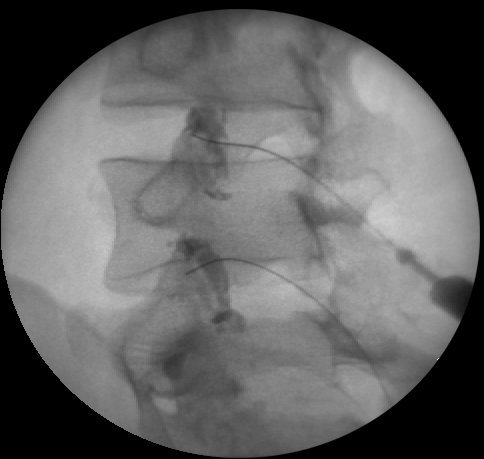
Diagnostic studies: Previous lumbar MRI. Negative SIJ block, negative discograms at L3/4 and L4/5, negative facet injections at L4/5 and L5/S1. Double diagnostic injections confirmed the painful facet joint at L3/4. Figures 1 and 2 show the facet joint arthrograms for this patient.
This case does not meet Revel=s criteria as the patient is under age 65 years, has worsening of pain with extension and exacerbation of pain with coughing. The SIJ pain provocation tests are all false positives. Presence of symptoms above the level of L5 helps to identify that the sacroiliac joint is not involved. There is no evidence of disc involvement based on pain behavior or movement loss with mechanical testing. The two positive tests in the hip examination are not helpful in explaining why the patient has constant low back pain. The presence of illness behavior helps to confound the situation, but it is important to note that there is an underlying pathology as determined by diagnostic injection. The mechanical assessment in this case can only be that the patient has a mechanically indeterminate source of lumbar pain.
Case #4: Symptomatic right L4/5 facet joint
50 year old female who related a one and one-half year history of constant right gluteal pain which commenced as a result of bending to wash her dog. She had recent onset of intermittent right lower extremity pain to the heel. Her pain was worsened by bending, sitting, prolonged standing, prolonged walking and was worse in the evening. She was better when lying in flexion. Her pain was not aggravated by coughing.
Movement loss: moderate loss of flexion and right sidegliding, major loss of extension
Test movements: Prior to testing: right lumbogluteal pain
RFIS no effect
EIS no change in movement loss following RFIS
REIS no effect
RFIL increased pain at end range, not worsened
REIL increased pain at end range, not worsened
RSGIS (R) decreased pain, no better
RSGIS (L) decreased pain, no better
SIJ tests: positive for compression, (R) Gaenslen=s, Patrick=s test was also positive.
Hip exam: all negative
Other: positive Spring test at L5 only
Diagnostic studies: Previous lumbar myelogram/CT. Lumbar MRI x2: Grossly arthrotic joint at L4/5, DDD at L3/4 and L4/5. Pt had a negative right SIJ injection and negative discograms at L3/4 and L4/5. Single diagnostic injection of the right L4/5 facet joint was provocation and analgesic positive.
Revel=s criteria are met in this case. The patient was better lying, had no pain exacerbation by coughing, and was not worsened by flexion, rising from flexion or extension. The sacroiliac joint can be ruled out as a pain generator as there are only two positive provocation tests. Patrick’s (FABERE’s) test is not a sensitive test for sacroiliac joint pathology.11 The lack of centralization of pain and lack of obstruction to movement with mechanical testing excludes the disc as a pain generator.
Case #5: Symptomatic right L5/S1 facet joint
48 year old male with a 16 month history of right sided low back pain which was worst with standing and was also worse with bending, rising from sitting and walking. Pain was relieved by sitting and side lying. There was no increase in pain with coughing.
Movement loss: major loss of extension
Test movements: Prior to testing: right PSIS area pain
RFIS no effect
EIS no change in movement loss
REIS increased pain at the PSIS, not worsened
RFIL not tested
REIL not tested
Extension with right rotation markedly increased pain, not worsened
Extension with left rotation NE
SIJ tests: positive for compression only
Hip exam: negative
Diagnostic studies: Previous lumbar MRI and myelogram/CT. Negative right SIJ injection, negative discogram at L5/S1. Double diagnostic injections confirmed the right L5/S1 facet as the pain generator.
Case #5 meets Revel=s criteria. This patient is the easiest to sort out of the five cases given. There is no peripheralization of symptoms and no evidence of pain stemming from lumbar discs, the sacroiliac joint or the hip.
Three of the five cases presented had double (confirmatory) blocks, which rules out false positive responses for the diagnostic facet injections. The remaining two have had negative diagnostic injections of adjacent structures, minimizing the likelihood of false positive facet injections. At least five out of seven of Revel=s characteristics are present in three of the cases presented, which would have correctly predicted the positive response to facet joint injection. The two cases in which less than five of the characteristics were noted were at the levels of L1/2,2/3 and L3/4. Revel=s study included only the lower lumbar levels, so it is possible that the characteristics which were identified may not apply to the upper lumbar spine. This would limit their diagnostic utility. It appears that Revel and colleagues may be justified in cautioning against using their facet joint characteristics for diagnostic purposes.
Discussion
There are a number of factors which confound the mechanical assessment of patients who have painful facet joints. Employing data collected from a larger group of patients who had positive diagnostic injections to establish the pain generator, the following factors were noted which can confuse the examiner:
1. Pain from chronically painful facet joints may be reported as being felt only at the PSIS. There may be no pain above the level of L5 to suggest lumbar involvement rather than the sacroiliac joint.
2. The upper lumbar facet joints can refer to the leg.
3. SIJ pain provocation tests may be positive in the presence of only lumbar pathology.
4. Lower extremity pain and paresthesias may be constant or intermittent, and there may be groin pain. Lumbar discs and sacroiliac joints can also be the source of pain and paresthesias in these areas.
What, then, are common characteristics of the dynamic mechanical lumbar assessment that may suggest symptomatic facet joints? The following list has been developed:
1. The pain may be bilateral or unilateral, but is never only in the midline of the spine.
2. There will be lumbar movement loss, but there is no change in the degree of movement loss with repeated movement testing.
3. There is no centralization or peripheralization with repeated sagittal plane movements.
5. The pain may be decreased or abolished with sagittal or rotational movements, but will not remain better as a result. Distal pain which simply abolishes without centralizing is not discogenic.
While this list is not definitive, it is a start toward identifying when the facet joint is the pain
generator. Additional research is needed to assess the ability of a dynamic mechanical evaluation to predict the presence of symptomatic facet joints. Until then, these patients remain mechanically indeterminate.
References
1. Donelson R, Aprill C, Medcalf R, Grant W. A prospective study of centralization of lumbar and referred pain. Spine 1997; 22: 1115-1122.
2. Helbig T, Lee CK. The lumbar facet syndrome. Spine 1988; 13: 61-64.
3. Jackson RP, Jacobs RR, Montesano PX. Facet joint injection in low-back pain. Spine 1988; 13:966-971.
4. Laslett M, Williams M. The reliability of selected pain provocation tests for sacroiliac joint pathology. Spine 1994; 1243-1249.
5. McCall IW, Park, WM, O=Brien, JP. Induced pain referral from posterior lumbar elements in normal subjects. Spine 1979; 4: 441-446.
6. Revel M, Listrat VM, Chevalier X, et al. Facet joint block for low back pain: identifying predictors of a good response. Arch Phys Med Rehab 1992; 73: 824-828.
7. Revel M, Poiraudeau MD, Auleley GR, et al. Capacity of the clinical picture to characterize low back pain relieved by facet joint anesthesia. Spine 1998; 18:1972-1977.
8. Schwarzer AC, Aprill CN, Derby R, et al. The relative contributions of the disc and zygapophyseal joint in chronic low back pain. Spine 1994; 19:801-806.
9. Schwarzer AC, Aprill CN, Derby R, et al. Clinical features of patients with pain stemming from the lumbar zygapophyseal joints. Is the lumbar facet syndrome a clinical entity? Spine 1994b; 19: 1132-1137.
10. Schwarzer AC, Aprill CN, Derby R, et al. The false-positive rate of uncontrolled diagnostic blocks of the lumbar zygapophyseal joints. Pain 1994c; 58: 195-200.
11. Young, S. Unpublished data.
12. Young S, Laslett M, Aprill C, Kelly C. The sacroiliac joint: a study comparing diagnosis by physical examination and contrast enhanced diagnostic block arthrography. Presented at the North American McKenzie Conference, New Orleans, 1998.
Figure 1. Case #3: Left posterior oblique and prone views of left L3/4 and L4/5 facet joint arthrograms.
These joints had positive provocation and analgesic responses with intra-articular injections. Positive responses
to left medial branch blocks of L2, L3 and L4 confirmed the diagnosis. (Courtesy of Charles Aprill, MD)